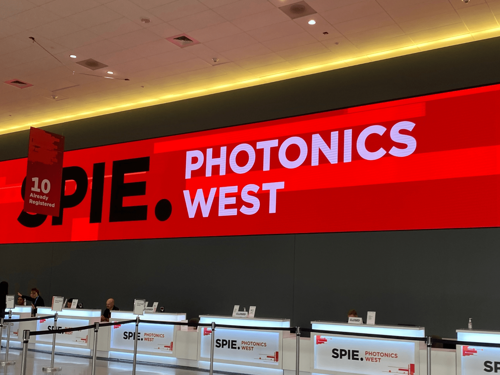
SPIE Photonics West 2026
RGB Lasersystems GmbH had the pleasure of exhibiting at this year’s SPIE Photonics West 2026. Over the course of the event, we welcomed numerous visitors to our booth and engaged in valuable conversations with long-standing partners, industry colleagues, and new connections.
We sincerely thank everyone who visited us and contributed to this successful experience. We look forward to building on these connections and turning shared ideas into future collaborations.

Laser World of Photonics 2025 Munich
We thank everyone who visited us and look forward to continuing these inspiring conversations.
Asia Photonics Expo 2025 Singapore
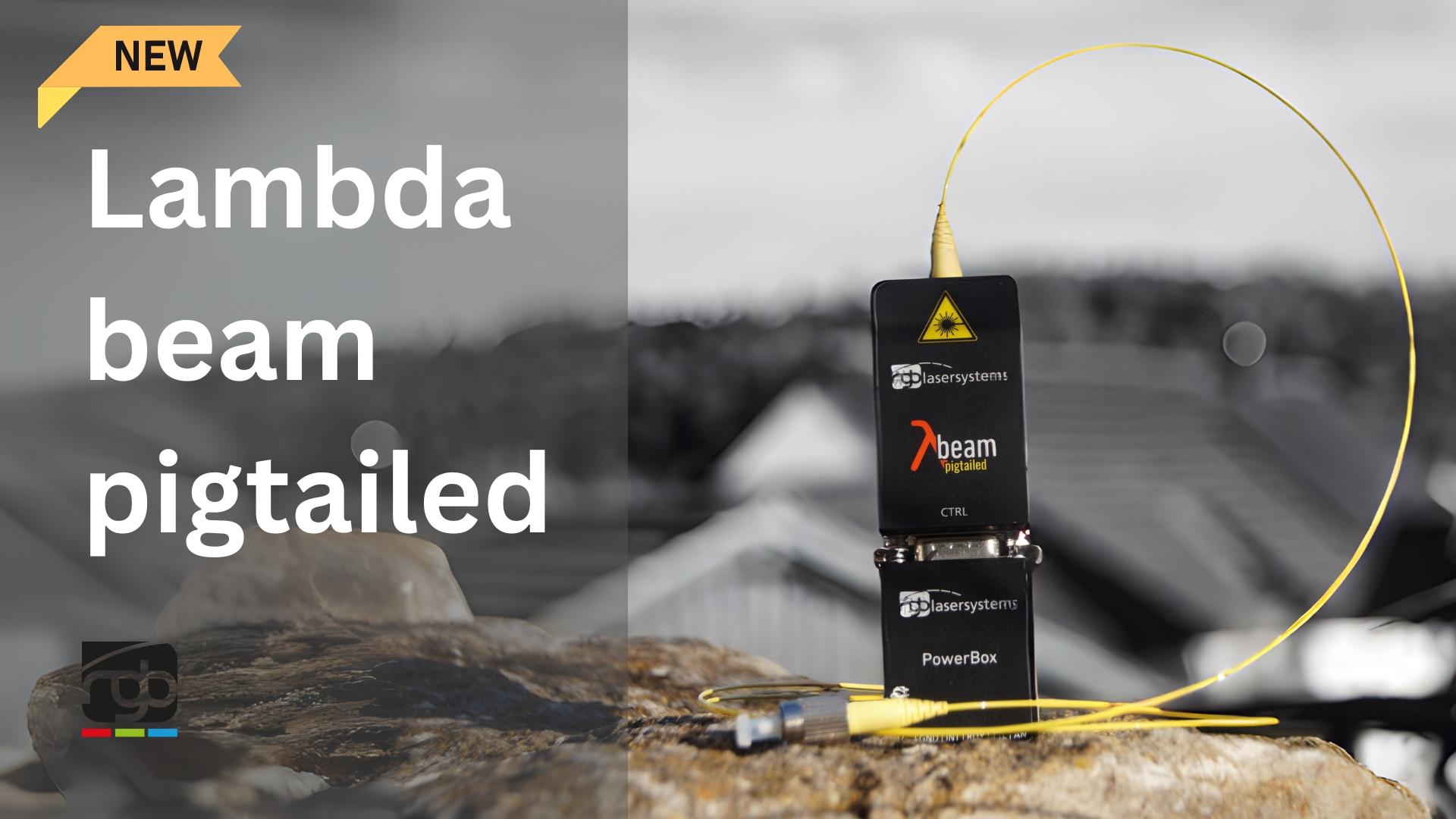
New product: Lamda Beam Pigtailed
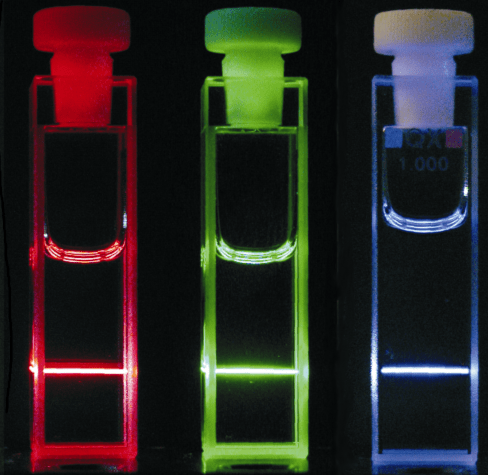
Spie Photonics West’24 San Francisco
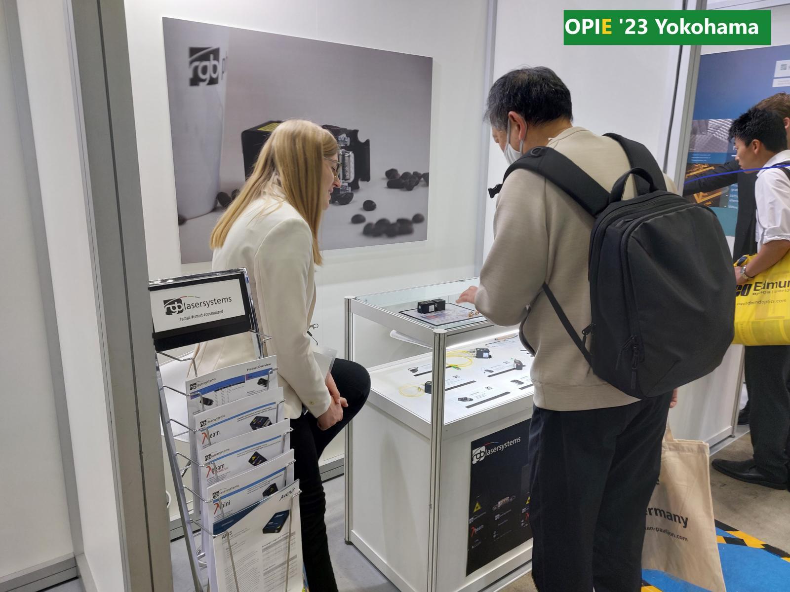
OPIE’23 Yokohama
Following applications also here on Linkedin
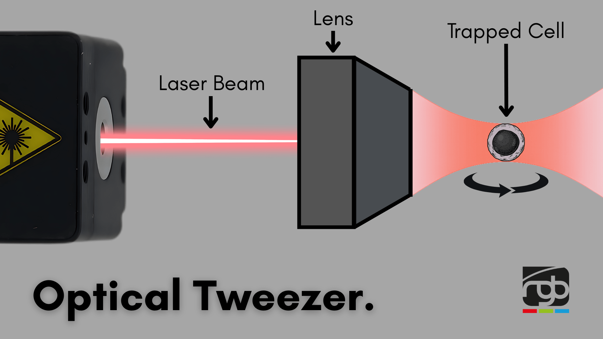
Optical Tweezers
What if you could hold and rotate a living cell using nothing but light?
Optical tweezers make that possible. By tightly focusing a low-power infrared laser (<1 W, typically 1064 nm), researchers can create a light-based force field that traps and manipulates microscopic objects like cells, bacteria, or even single molecules – entirely without physical contact.
How it works:
The laser creates a steep intensity gradient that pulls small particles into the beam’s waist. Using dual beams or beam shaping, it’s even possible to rotate objects in 3D.
Why rotate a cell?
Because in cell biomechanics, drug delivery research, or 3D imaging, orientation matters. Scientists need to examine how a cell behaves when force or stimuli are applied from specific directions – revealing things that static views would miss.This breakthrough was so impactful that it earned a Nobel Prize in Physics in 2018 – proving how a simple beam of light can act as a precise tool for biological manipulation.
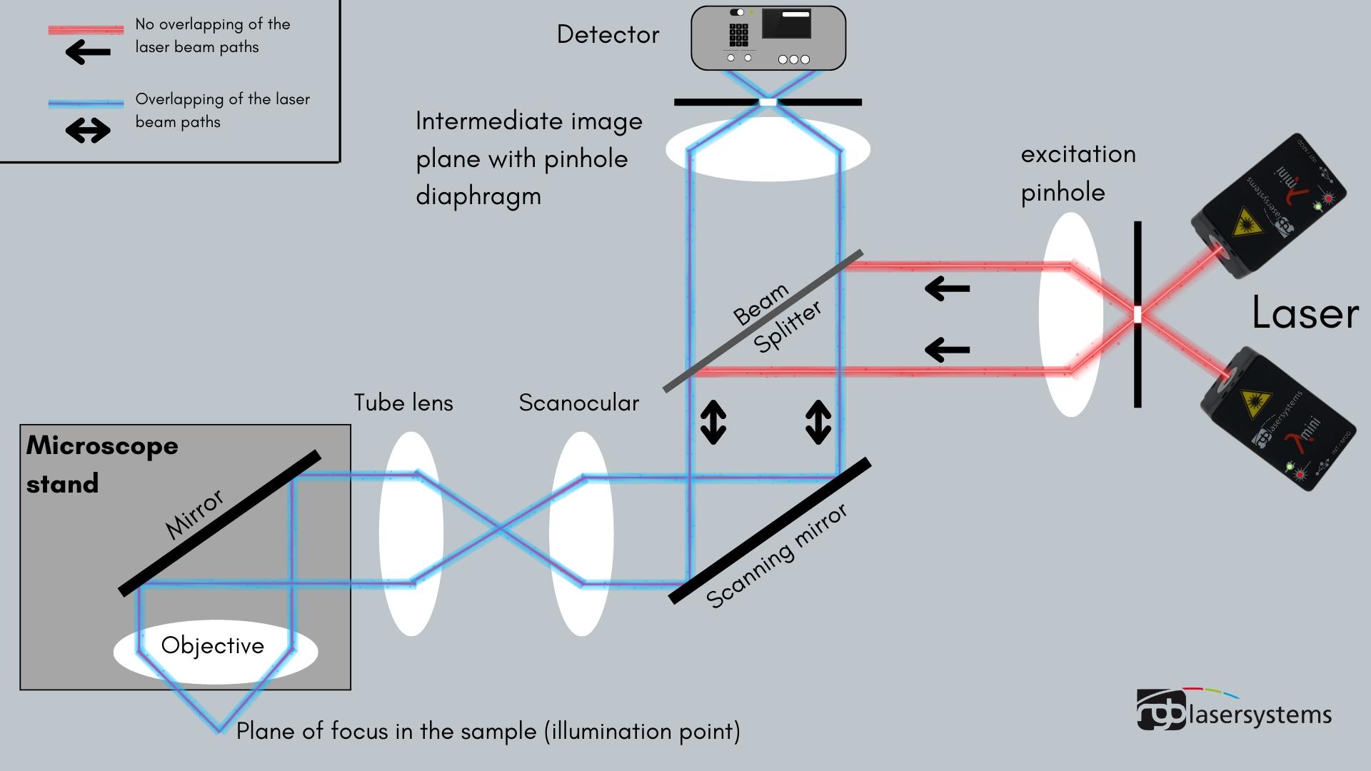
Confocal microscopy
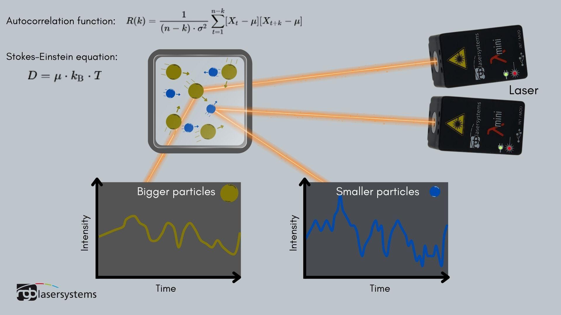
Dynamic light scattering/photocorrelation spectroscopy
This is a measuring method that can be used to determine the hydrodynamic radius of particles.
When particles (e.g. proteins) are radiated by a laser light, which is coherent and monochromatic, it is scattered in all directions.
With the help of an interferometer, the distance between centers from which the scattered light originates (so-called scattering centers), i.e. the distance between particles, can be determined.
As the distances between the particles are constantly changing due to Brownian molecular motion (random movements of molecules/particles in a fluid state), interference occurs which leads to fluctuations (changes in scattering intensity). The diagram shows what such fluctuations look like graphically for large/small particles. The interference can now be used to determine the speed of the particles and the so-called diffusion coefficient. The autocorrelation function can be used to calculate particle parameters, which can be used to determine the hydrodynamic radius of the particles using the Stokes-Einstein equation. These results can be used to indirectly determine the molar mass of the measured particles.
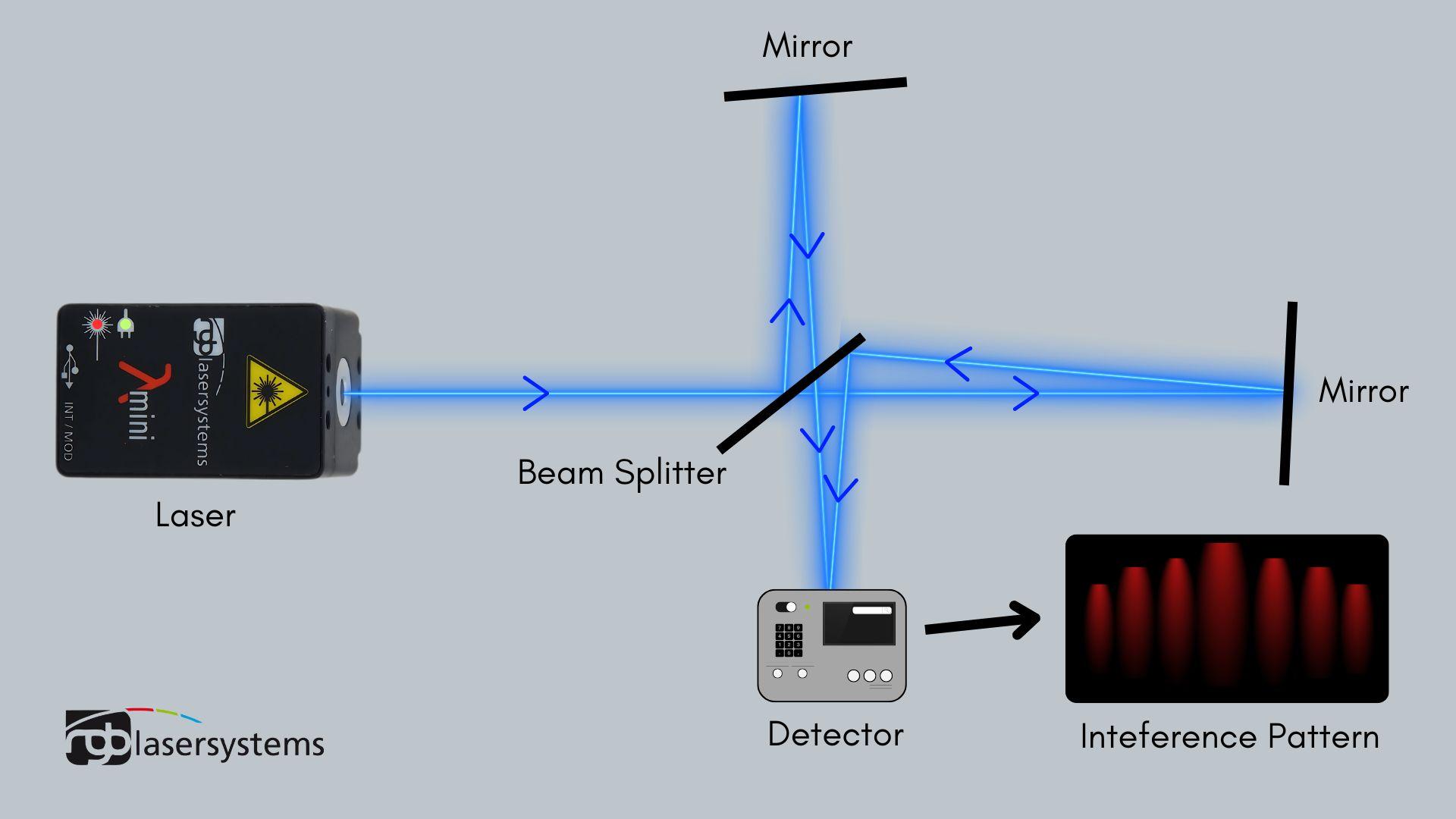
Michelson Interferometer
The Michelson interferometer utilizes the phenomenon of interference, which can only be observed with coherent light, for example that of a laser.
The graphic illustrates the basic structure of the Michelson interferometer. An interferometer splits a light wave into two parts. These two waves then travel different distances with different propagation times. When they meet, interference occurs.
If the optical path length of one of the two waves is changed, e.g. by moving one of the two mirrors, the phases of the two waves are shifted against each other and thus the interference pattern changes.
By measuring the intensity of the resulting wave, even the smallest changes in the path difference between the two waves can be measured.
One example of an application is the measurement of the thermal expansion of materials. Using the Michelson interferometer, even the slightest expansion can be detected, making the measurement extremely accurate.
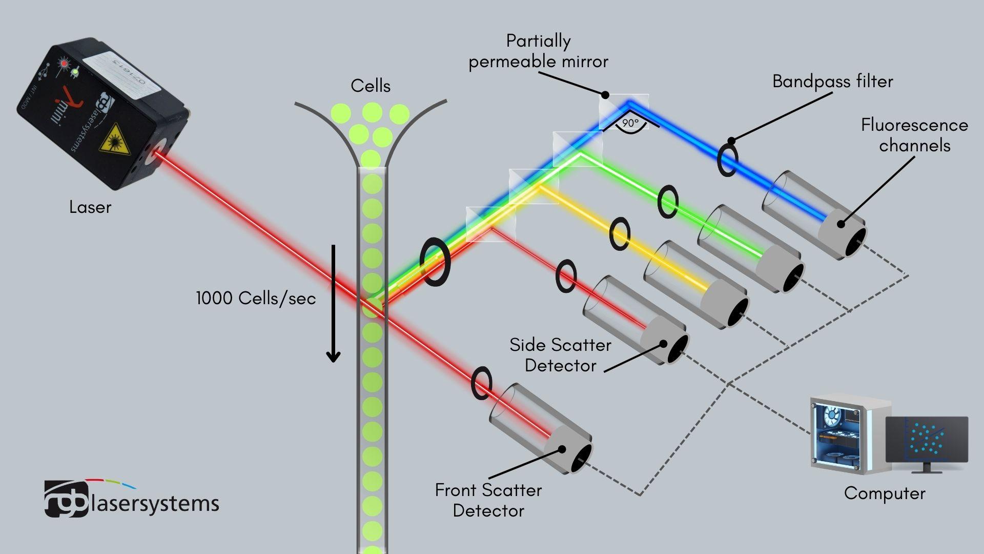
Have you ever heard of flow cyometry?
This is a measurement method for the qualitative and quantitative analysis of cells. It starts with cells being sucked into a fluidic system at high speed. The more than 1000 cells per second are shot individually through the laser beam and emit fluorescence depending on their individual characteristics. With different wavelengths (red, yellow, green, blue…) the following different information can be obtained. The forward scatter detector is a measure of the diffraction of the light, which can be used to determine the volume of the cell. The side scatter detector is a measure of the refraction of light at right angles, which can be used to determine the size and structure of the cell nucleus, the number of vesicles and the granularity of the cell. Using a so-called FACS- (Flourescence-activated Cell Sorting) device, the cells can then be separated according to their characteristics.
One example of an application of flow cyometry is the separation of sperm cells with sex chromosome X and those with chromosome Y in order to determine the sex of an embryo created by in vitro fertilisation.
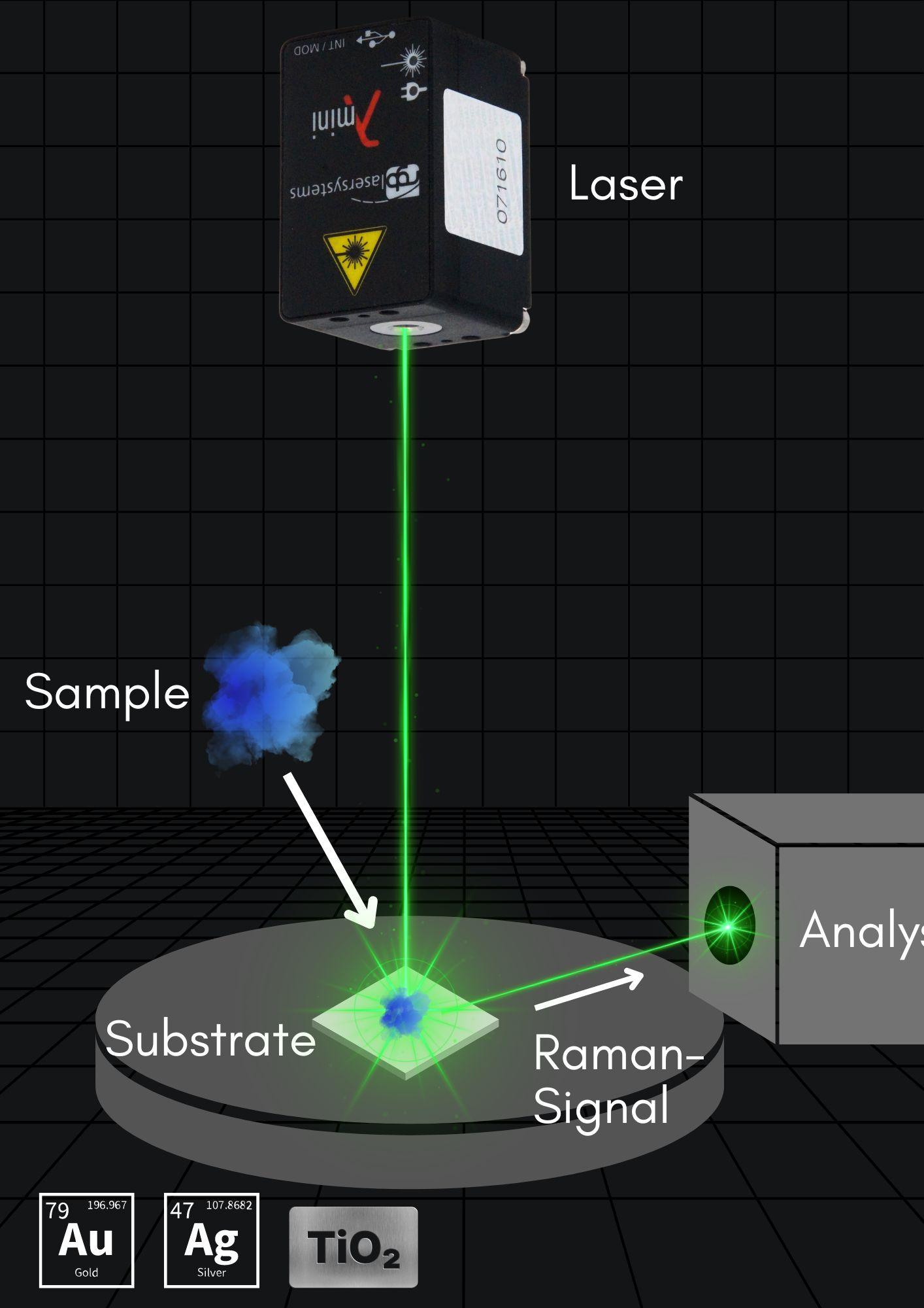
Developments in Raman Spectroscopy
Until now, expensive and often unreliable methods such as the SERS- (surface-enhanced Raman scattering) method have been used for ultrasensitive analyses.
A research team from the field of materials science has succeeded in developing a method that should increase performance 50-fold.
A Raman measurement works as shown simplified in the picture. The laser plays a decisive role here, as does the substrate onto which the sample gets placed on.
The team has developed a substrate made of different materials with different characteristics, which together can have a positive effect.
For example, it contains four plasmonic nanostructures, including gold and silver particles, which generate a light-matter interaction.
These interactions are also known as plasmonic effects. Combined with a photocatalytic titanium-oxide-layer to cause a photocatalytic effect, the RAMAN signal is intensified by a factor of 50.
This ensures that the sample can be analysed much more precisely and quickly.
A special feature of the substrate is that it can be cleaned using UV light and can be used up to 20 times, which reduces costs immensely.
Energy Transfer Upconversion (ETU)
Energy Transfer Upconversion is a physical principle that involves the excitation of a laser-active ion to a level above that which would be achieved by simple absorption of a pump photon, the required additional energy being transferred from another laser-active ion.
The Institute of Chemistry at the University of Basel uses a customized Lambda Beam laser system. By means of an individually arranged lens, the laser achieves a very high power density with only 40mw output power. Very interesting for Upconversion studies.
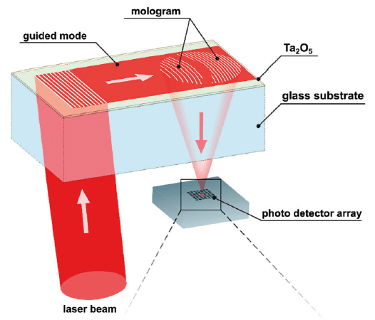
Ever heard of Molography?
In short, focal molography is a nanotechnology-based method that cleverly combines photolithography, molecular self-assembly and state-of-the-art optical technology.
Hereby a 785nm RGB laser light wave hits antibodies, part of the light is diffracted (redirected) at a certain angle, while the angle is defined by the spacing of the antibody lines.
The patented biosensor from lino Biotech AG evaluates the result and finds applications in quality control for cell and gene therapies, drug screening in living cells or point-of-care diagnostics in healthcare.